Please activate JavaScript!
Please install Adobe Flash Player, click here for download
ePaper created 2011-01-05, 11:59:18 | version 1.19.1
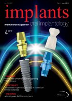
I user report _ oroantral communications Figs. 14a & b_Histological analysis of soft (left) and hard tissue (right). Harvesting and preparation artifacts are visible. notsatisfyingfromanestheticpointofview.Therefore, a membrane-based approach in combination with a method to preserve the alveolar ridge after tooth ex- traction is favored. The perforation can be closed with amembraneand/oratissuetransplantandtheextrac- tion wound is filled with a material suitable for socket preservation(Beckeretal.1987;Watzek2008).Finally, the wound is sealed towards the oral cavity with an- other membrane and/or soft tissue transplant. How- ever,thistechniqueislaborious,costlyandismastered mainlybyexperiencedoralsurgeons.Membrane-inde- pendent, minimally invasive subantral augmentation methodsaresimpleandefficientone-stepprocedures forclosureoforoantralcommunications(Thomaetal. 2006).Themethodpresentedinthiscase-cohortstudy critically depends on the unique features of the novel syntheticgraftmaterial.Initiallyitismoldable,andcan be inserted into defects of any shape. In contact with blood, the material hardens and forms an inherently stable but openly porous body. Most likely, coagulated blood in the interconnected pores and the underlying, stabilized coagulum supports the formation of an air- tightbarrier.Duringthefirstdays,thepolymerlayerthat mediatestheformationofastablescaffoldtakesupwa- ter, which results in a slight volume increase and guar- anteesatightcontactbetweenthedefectwallsandthe graft material. The -TCP composite is tissue-friendly and the phase-pure -TCP is degraded in parallel with bone formation (Nair et al. 2006; Rothamel et al. 2007; Gläser2009). Unlikeothermethods,thedescribedsub- antralaugmentationproceduredoesnotinflictfurther damagetothesoft-andhardtissuesurroundingthede- fect site. Pain, swelling and increased risk of infection thatresultsfromsofttissueandperiostslittingandthe exposure of the underlying bone are avoided, which representsamajorpatientbenefit.Ittooktwoweeksfor thegraftmaterialtobecoveredwithsofttissuewhereas defect closure is immediate with other techniques. It couldbearguedthatthedelayedcoveragewithsofttis- suemayincreasetheriskofinfection.However,neither in the present study nor in a larger study that used an identical, thermally molded -TCP composite for oroantral communication closure did such infections occur (Gacic et al. 2009; Thoma et al. 2009). Further studies will need to compare larger patient groups to address this issue in more detail. The clinical and radi- ographic controls showed an almost complete preser- vationofsoftandhardtissuestructuressimilartomem- brane-based surgical techniques. Expansion of the si- nus cavity towards the alveolar ridge was observed in onecase18monthsaftertoothextraction,makingasi- nusliftforstableimplantplacementnecessary(Fig.12). The -TCP composite is replaced by bone that is sub- jected to physiological bone remodeling. Expansion of the sinus cavity thus occurred like it would be the case inanynon-augmentededentuloussetting. The described minimally invasive method for oroantral communication closure is well suited for oroantral communications that are located above un- problematic extraction sites. However, it should not be appliedifthesizeorthegeometryofthedefectdoesnot allow stable anchorage of the material. Alternative methods should be considered in patients with very large oroantral communications that may result from theextractionoftwoormoremolars.Insuchcases,the graft material may be lost or displaced into the sinus cavitybymasticationforces.Also,safeanchorageofthe materialandefficientclosureoftheoroantralcommu- nicationmaynotbegiveninshallowextractionsockets, e.g. if the buccal lamella is missing. A perforation with only3to5mmremainingverticalboneheightcouldbe successfully closed during this study. This patient was monitoredmorecloselythandescribedintheprotocol. Defects shallower than this certainly should be treated withanalternativemethod. In conclusion, the described minimally invasive method for closure of oroantral communications has several obvious advantages over conventional tech- niques.Itisfastandefficientanddoesnotinflictdam- age to the surrounding tissue since no additional sur- gery is needed. Nevertheless, alternative methods shouldbeconsideredifanunsuitabledefectgeometry jeopardizes stable graft anchorage. Soft and hard tis- sue structures were mostly preserved, and no compli- cations or failures to close the perforations were recordedduringthisretrospectivecasecohortstudy._ Article scheduled for publication, based on the article “Der Einsatz von polylactid-beschichtetem -Tricalciumphosphat zum MAV-Ver- schluss”,publishedinQuintessenz08. 36 I implants4_2010 Fig. 14bFig. 14a DrStefanNeumeyer DrStefanieNeumeyer-Wühr Lemminger Straße 10 93458 Eschlkam,Germany E-mail: praxis@dres-neumeyer.de _contact implants